The ESPript interface consists of:
- A BUTTONS frame, fixed at the top of the page.
- A MAIN frame, which contains the user form.
- A POP-UP window containing the results of your ESPript job.
- Fill up the form by uploading at least one multiple alignment file in the Aligned Sequences .
- PDB, CIF or DSSP files can be uploaded in the Secondary structure depiction section.
- The rest of the form allows you to change parameters related to the secondary structures and sequence similarity depiction, as well as the layout of the alignment output or the size and format of the resulting figures (PostScript, PDF, PNG, TIFF).
- All these options are explained in detail in the User Guide section or directly from the interface by clicking on the
 icon. icon.
- Tooltips are also available for form elements by moving the cursor over an
 Answer to the Ultimate Question of Life, the Universe, and Everything Answer to the Ultimate Question of Life, the Universe, and Everything

Default: 42 icon.

- Only yellow buttons are activen with the exception of the TIME bar. Blue buttons are not clickable.
- When the main form is filled in, click on the SUBMIT button to let ESPript process your query.
- A result pop-up window will automatically appear within seconds. This results window can be (re-)opened at any time by clicking on the RESULTS button.
- In order to access the results, you may need to authorize your browser to display pop-up windows from
espript.ibcp.fr.
If necessary, consult your browser documentation.
- The DOC button displays the full User Guide in a separate window.
- Click on ADV (ADVanced) or EXP (EXPert) to have access to more options. The default mode is BEG (BEGinner).
- In ADV mode, you can import another secondary structure file, change secondary elements labels, tinker with special characters and calculate similarity scores between groups of sequences.
- In EXP mode, you can also define your own colors, shift sequence numbering, etc.
- You can navigate between BEG , ADV and EXP modes without losing any information in your query.
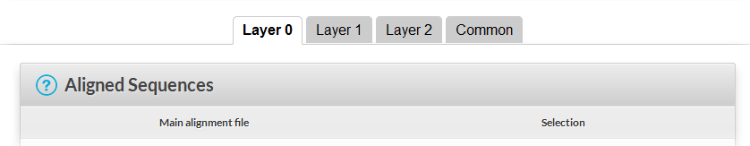
- If you are in ADV or EXP mode, you can use the +1 button to build a layered ESPript figure. When you click on +1 , a new layer is created (up to 20 layers can be defined). The parameters set for Layer 0 (i.e. the first Layer) are copied to Layer 1 (and so on). Button -1 allows the user to suppress the last layer created. Layer Common contains parameters that are common to all layers.
- You can switch between the different layers by clicking on the tab bar (see below). See example #3 to learn more about this option.
- Use the SAVE button to save your session to your computer, so that you can use the same parameters and files later.
- Use the LOAD button to load a previously saved session.
- Pay attention to the TIME bar. You must execute at least one command every 60 minutes otherwise your session will be closed.
- Before leaving, click on the EXIT button to permanently remove the processing and result files from the server.
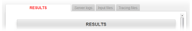
The RESULTS pop-up window:
- Figures produced by ESPript appear in this multi-tab window with dedicated links. Left-click to visualise them, or right-click to retrieve them.
- Expert (or curious!) users can click on the Server logs Input files or Tracing files tabs to access the corresponding reports and data.
EXAMPLE #1 - Beginner Mode
Session vp7_beg.sav for beginners (resulting PDF, PostScript, PNG).
Sequences homologous to vp7_btv10, a viral protein whose 3D structure has been solved, have been aligned using ClustalW.
The session file vp7_beg.sav allows the user to create a figure from the alignment and to display secondary structure elements of vp7_btv10. It has been created as follows:
- Start ESPript.
- Save the alignment file
vp7.aln on your disk, and upload it in the first section Aligned Sequences of the form.
- SUBMIT and open the resulting image. A column is framed in blue if more than 70% of its residues are similar according to physico-chemical properties (threshold is set to 0.7 in the Sequence similarities depiction parameters section of the form and the button %Equivalent is activated). Note that
vp7_btv10 is the second sequence in the alignment.
- Save the PDB file
vp7_btv10.pdb on your disk and upload it in the second section Secondary structure depiction of the form.
- SUBMIT and open the resulting image. Note that the secondary structure elements displayed above the sequences block are named
vp7_btv1s. This is normal: by default they are aligned to the first sequence displayed by ESPript, here vp7_btv1s. However, this is wrong: your secondary structure elements refer to vp7_btv10.
- Scroll down the form to the Defining groups section. Change the string
all to 2 all.
- SUBMIT and open the resulting image.
vp7_btv10 is now the first displayed sequence and secondary structure elements are correctly aligned with it.
- Check the Alignments output layout section. You will see a yellow tooltip stating "
Suggested number of columns: 60 (for A4, portrait and a font size of 7)". This indicates the Number of columns to set in order to obtain a justified figure.
- SUBMIT and SAVE the session.
EXAMPLE #2 - Advanced Mode
Session vp7_adv.sav for advanced users (resulting PDF, PostScript, PNG).
This session uses the same ClustalW alignment as in Example #1. You can start with the vp7_beg.sav session described above.
- Start ESPript.
- LOAD the previous session by using the buttons frame.
- SUBMIT to check that everything is OK.
- Click on ADV to access new options.
- Tick the button Relative accessibility in the Secondary structure depiction section of the form, to show accessibility per residue with a blue bar below the sequences block on the image.
- Click on Hide names in the same section, to remove the string
vp7_btv10 above the sequence blocks, on the same line as the secondary structure elements.
- SUBMIT to check.
- Type the command
U R 127 250 in the Special commands and characters section, to add two red triangles below residues 127 and 250 of the first displayed sequence, vp7_btv10. These triangles show the separation between an α-domain (1-127, 250-349) and a β-domain (128-249) in the 3D structure.
- SUBMIT to check.
- In the same section, type
S B 168-170 178-180 to mark an RGD tripeptide observed at different positions in the sequences. Then type:
X B 1-126 254-349 to color secondary structure elements of the α-domain in blue.X G 127-253 to color secondary structure elements of the β-domain in green.T R 1 2 to colour the names of the first and second sequences displayed in red.- Note: you must press [ENTER key] after each line of special commands.
- SUBMIT to check.
- Type in Footnotes subsection of Alignments output layout the sentence:
Alignment for protein vp7.
- SUBMIT to check.
- In the Defining groups section, delete
2 all and type 2 1 3-6 7-8 [ENTER key] 9 in order to define three groups of sequences according to phylogeny (the first group consists only of btv sequences) and to calculate in-group as well as cross-group similarities. This necessits the use of a scoring matrix, and the option Risler is selected instead of %Equivalent in the Sequence similarities depiction parameters section.
- SUBMIT to check. You can now observe:
- In column 1: that all residues are identical and are boxed in red.
- In column 10: that residues of the first group are identical and are in red (all threonines) and that those of the second group are similar (threonine and serine) and are in red ; the third group consists of a single sequence and the residue is in black. The global similarity score calculated from all groups is > 0.7 (which is the threshold entered in the Similarity Calculation section) and the column is framed in blue.
- In column 19: that residues are not framed, the global similarity score being < 0.7. Moreover, residues of the second group are in black, because they are not similar (lysine and threonine) according to a in-groups score < 0.7 (the same threshold is used by the program for global scores and in-groups scores).
- In column 79: that the column has a yellow background. Residues are similar in each group (in-groups scores > 0.7) but significantly different from the first group (all prolines) to the second (all histidines). Thus, the cross-groups score is > 0.5, which is the current threshold according to the Sequence similarities depiction parameters section.
- Remark: try this if you want a more colorful figure:
- Select the Thermal option in the Alignments output layout section to obtain bold characters.
- Type the command
M Y all in the Special commands and characters section to add a yellow background to similar residues.
- SUBMIT to check.
Session vp7_exp.sav for experts (resulting PDF, PostScript, PNG).
Again, the same CLustalW alignment, but now with information on the 3D structure of vp7_btv1s. The button +1 of the buttons frame is used for this purpose. The following needs to be generated in ADV or EXP mode. We will start from scratch.
- Open ESPript.
- Upload
vp7.aln in the Aligned Sequences section.
- Upload
vp7_btv10.pdb in the Secondary structure depiction section.
- Type
X B 1 in the Special commands and characters section, to color in blue secondary structure elements and align them with the first displayed sequence.
- Type
h in the Replace secondary structures labels subsection of Special commands and characters , in order to remove the label h1 from the 310-helix.
- Change
all to 2 all in the Defining groups section to display vp7_btv10 as first sequence in the alignment.
- SUBMIT to check the result.
- Click on Hide sequences in the first section and SUBMIT again. Only secondary structures elements are now displayed.
- Click on +1 in the buttons frame to create a new layer.
- The form now contains two independant layers named Layer 0 and Layer 1 and a shared one ( Common ). Check that the alignment file
vp7.aln has been copied from Layer 0 to Layer 1 .
→ All following commands must be entered in Layer 1 .
- Disable option Hide sequences , then enter a Vertical shift of -1 in the Alignments output layout section.
- SUBMIT to check. You now have a gap between secondary structure elements of
vp7_btv10 and sequences.
- Upload the
vp7_btv1.dssp file in the TOP secondary structures section of the box Secondary structure depiction , which corresponds to vp7_btv1s.
- Type
X R 2 in the Special commands and characters section, to color in red secondary structure elements and align them with the second displayed sequence.
- Type
2 all in the Defining groups section.
- SUBMIT to check.
→ Information on secondary structure elements are well aligned with sequences but labels of vp7_btv1s need to be removed.
- Click on Hide labels in the Secondary Structure section.
- SUBMIT to check.
→ All information is now well displayed. We will add extra boxing on residues.
- Type
Q P 1-3 169-171 in the Special commands and characters section. This means that columns 169-171 of sequences 1-3 are boxed in pink. Note that the program uses columns numbering instead of residues numbering for the special commands Q, V and W. You can visualize columns numbers by checking the button Ruler in the same section.
- Type
Q P 5-7 169-171 [ENTER key] Q P 8 180-182 in the same section to box all RGD tripeptides.
- Select the Flashy mode in the Color scheme section of the Common tab to add a yellow background on similar residues.
- SUBMIT to check.
→ We will now color sequence names of vp7_btv10 and vp7_btv1s. Type in the same Special command and characters section.
T B 1 to color the name of the first displayed sequence in blue.
T R 2 to color the name of the second displayed sequence in red.
→ Finally, we will number all sequences.
- Click on Number sequences in the Aligned Sequences section of Layer 0 .
- SUBMIT to check.
|